Dr_c17_cavalheiro_s
CHAPTER 17Hydrocephalus in Neurocysticercosis and Other Parasitic
and Infectious Diseases
S. CAVALHEIRO*, S.T. ZYMBERG, P.D. TEIXEIRA, M.C. DA SILVA The cestode species are the most common parasites infection by T. solium. Colli et al. reported that 2.7% that affect the central nervous system (CNS). Five of hospital admissions for neurological diseases in different cestode infections of the nervous system the city of São Paulo in 1986 were due to neurocys- are: cysticercosis, from the larva of Taenia solium – ticercosis [17]. Admissions in the pediatric age the most common of them, with more than 50 000 group due to neurocysticercosis comprised 2%-10% people infected worldwide; hydatidosis (hydatic of all cases. The habit of repeatedly introducing cyst disease), from the larva of Echinococcus granu- their fingers into their mouths and easy contact losus; alveolar cyst disease from the larva of with the soil expose children to a higher risk of Echinococcus multilocularis; coenurosis, a rarer massive cestode infections. Statistically, older peo- type, from the larva of Taenia multiceps; and, ex- ple are less affected [12, 39].
ceptionally, sparganosis from the larva of Spirome- Cysticercosis affects mostly people with lower liv- tra mansonoides. Only the most important ones and ing standards. Though a few studies have shown a those related to hydrocephalus will be discussed higher incidence among males, there seems to be no here – cysticercosis and hydatidosis. Three other sex preponderance, and as to age, the peak incidence infections are common and may lead to hydro- is between 25 and 35 years [52].
cephalus. They are fungal, viral, and infections byMycobacterium tuberculosis. Other bacterial ventri-culitis will not be discussed in this chapter.
Incidence of Hydrocephalus in
Neurocysticercosis
In general terms, hydrocephalus is responsible for 15%-30% of clinical manifestations of neurocys-ticercosis [42]. Neurocysticercosis can be classifiedinto two forms: benign and malignant [25]. In the benign form, the parasites are usually parenchyma-tous, on the meninges outside the cisterns, or, in a Cysticercosis infects about 50 million people small number of cases, ventricular. Patients are around the world, 50 000 of whom die each year. It asymptomatic or present with headaches or is found, alongside pork tapeworm infestation, in seizures. Prognosis is good and response to treat- countries with poor hygiene habits. It is rare in ment is quick. In the malignant form of the disease, most of Europe; in Asia reports are sparse. It occurs the parasites are located in the subarachnoid space, frequently in India, the northern coast of Africa, es- inside the cisterns or inside the ventricles. Blood pecially Egypt, and among the natives of southern vessels are affected. Hydrocephalus is the most com- Africa. Cysticercosis is rare in the United States of mon clinical presentation and may be the result of America, but is the leading cause of hydrocephalus basal arachnoiditis or of the presence of intraven- and seizures among Hispanic Americans living in tricular cysticerci. Patients may present with myriad Los Angeles and California [4, 59]. Latin America is symptoms or develop symptoms of increased in- the area with the highest incidence of cysticercosis tracranial pressure. Prognosis is poor and therapeu- – it is referred from Mexico to Argentina and Chile.
tic response not good. Wei et al. [70] analyzed 1400 From all autopsies in Mexico in 1979, 1.9% showed cases of cerebral cysticercosis and detected ven- Department of Neurosurgery, Escola Paulista de Medicina, Federal University of São Paulo, Rua Dr. Oscar Monteirode Barros, 400/21, 05641-010 São Paulo, Brazil
Chapter 17 • Hydrocephalus in Neurocysticercosis and Other Parasitic and Infectious Diseases
triculomegaly in 204 patients (14.5%). Seventy pa-
Transmission and Life Cycle of the Parasite
tients had global dilatation of the ventricles and 67(32.8%) isolated dilatation of the fourth ventricle. In1984, Apuzzo et al. [4] demonstrated in a series of 45
Humans are the only definitive hosts of T. solium, har-
cases of intraventricular cysticercosis that the
boring the mature tapeworms in the lumen of the
fourth ventricle is most commonly affected (24 pa-
small intestine. Usually, there are only minimal clini-
tients). In 12 cases, the cysticerci were located in the
cal symptoms or even no symptoms at all. The mature
third ventricle, and in 5, inside the lateral ventricles.
T. solium is composed of a head or scolex that con-
This series, however, did not show any case of isolat-
tains four suckers and a double crown of hooks, a nar-
ed blockage of the temporal horn of the lateral ven-
row neck, and a body that is sometimes many meters
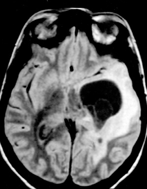
tricle. Conversely, in our series the presence of cysts
long and is composed of hermaphroditic proglottids.
completely blocking the temporal horn is somewhat
A few proglottids are released daily from the distal ex-
common (Fig. 1). The cysticerci may move from one
tremity of the tapeworm and eliminated with the
ventricular cavity to another, a fact that has been
host's feces. The host thus delivers proglottids with
verified during ventriculography [4, 15].
large numbers of fertile eggs to the exterior, wherethey contaminate the soil. These eggs are resistant tobeing dried out and may remain in the soil or waterfor months. In some cases, the eggs are released insidethe intestinal lumen, and these are the eggs releasedwith the host's feces. Around 50% of these eggs are
mature and fertile. The usual intermediate host, thepig, becomes infected by ingesting water or food con-taminated by human feces. The parasites then takeshelter in organs with a high oxygenation and move-ment level, such as the brain, chewing muscles,tongue, and heart (Fig. 2). By consuming uncookedpork containing the larva (intermediate form), hu-mans acquire tapeworms – the mature form of theparasite (Fig. 2).
Human cysticercosis may result from two mecha-
nisms: autoinfestation and heteroinfestation. In theformer, the proglottids may release the fertile eggsinside the host's intestines, leading to internal self-in-festation. This is considered one of the ways man canbecome infected by T. solium. In order to liberate thelarvae, the eggs need to be exposed to the stomach
acid. Thus, vomiting or antiperistaltic movements ofthe intestines push proglottids with fertile eggs to-ward the stomach. These proglottids may, in turn,fragment and release their eggs to the effects of thestomach acids, and suffer, back into the duodenum,intestinal digestion and disintegration of the em-bryophore and release of the oncosphere. In thesmall intestine, the larva attaches itself to the intesti-nal wall using its hooks, and insinuates itself betweenthe epithelial cells of the villi, with the help of cy-tolytic substances secreted by special glands. Itreaches the connective tissue corium and, once thelumen of the capillaries is reached, invades the tis-sues through the blood vessels. Other facts corrobo-rate this infestation mode, such as the presence inone individual of both cysticerci and the maturetapeworm. A history of tapeworm infection is found
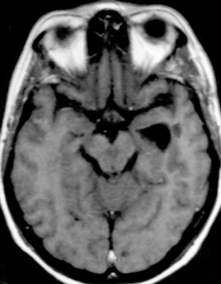
Fig. 1. a Axial FLAIR MRI demonstrating blockage of the
temporal horn by cysts. b Axial T1-weighted MRI after en-
in 7%-22% of patients with cysticercosis [59]. Exter-
doscopic cyst removal
nal self-infestation occurs when the individuals are
Fig. 2. Life cycle of the parasite
contaminated by their own feces, ingesting eggs or
blood stream. The stomach is thus the first barrier
proglottids of their own tapeworm [2].
against this infestation, as only eggs whose shells
The mechanisms involved in heteroinfestation in-
have resisted the gastric acidity will release their
clude ingestion of water, vegetables, or fruit contami-
oncosphere in the intestinal alkaline medium. Once
nated by T. solium eggs, either through poor hygiene
liberated in the blood stream, the embryos will be
habits or by soil fertilized with human feces. Usually,
retained in places where the diameter of the vessels
consumption of contaminated pork leads to tape-
is too small or the circulation slow (muscle, retina
worm infection. However, immune-suppressed indi-
viduals may become infected by contaminated pork
The larval form of T. solium, Cysticercus cellu-
containing larvae.
losae, develops from the embryo. It possesses a
The mature proglottids of the T. solium are elim-
head and a neck, invaginated inside a vesicle. The
inated from the intestine with the feces into the en-
head is similar to that of the adult form and the
vironment, degenerate, and release the eggs (em-
neck is very small; both are wrapped around them-
bryophores), which harbor the embryo (hexa-
selves. The vesicle is clear, semitransparent, of vari-
canth). Once ingested, the egg undergoes the effects
able shape, and measures about 10-15 mm; its inte-
of the gastric acids and passes to the intestines
rior is filled with crystalline fluid. When the cys-
where, 24-72 h afterwards, its shell fragments, re-
ticercus is located within the cerebral ventricles or
leasing the oncosphere. The oncosphere, in turn,
in the subarachnoid space around the brainstem,
penetrates the intestinal wall with the help of its
the head and neck may disappear and secondary
hooks, reaches the mesenteric veins and then the
vesicles may form from its walls, mostly intercon-
Chapter 17 • Hydrocephalus in Neurocysticercosis and Other Parasitic and Infectious Diseases
nected, taking the aspect of an irregular grape clus-
located preferentially at the level of the basal gan-
ter (2-15 cm). This cluster is called Cysticercus
glia and cerebral cortex due to their increased vas-
racemosus [13, 14].
Once implanted in the CNS, the hexacanth begins
its development onto the embryonic form, the cys-
Pathological Findings in Neurocysticercosis
ticercus [24]. In its first stage of development, calledthe vesicular stage, the membrane is thin and trans-
From one to hundreds of embryos may reach the
parent, the fluid is clear, and an invaginated scolex is
CNS, where they may survive for 1-30 years in the
the norm. The cysticerci can remain in this phase or
form of Cysticercus cellulosae, or as Cysticercus race-
start a degenerative process as a result of the host's
mosus (its intermediate form) or even as both forms
immune response, which may lead to their destruc-
coexisting together. Once settled, the cysticerci, while
tion in three stages. The first stage of this process,
alive, cause almost no response from the surround-
when the vesicular fluid becomes cloudy and the
ing tissues. The inflammatory response is related to
scolex shows signs of hyalin degeneration, is called
the number of parasites and their state of degenera-
the colloidal stage. Later on, the walls of the cyst be-
tion, which, in turn, depends essentially on the re-
come thick and the scolex is transformed into a gran-
lease of antigens [26]. In many cases, the immune re-
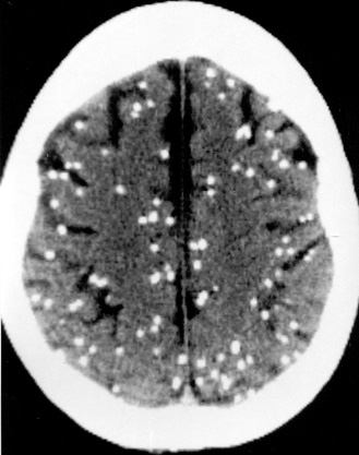
ular mineralized structure. Lastly, the whole parasite
sponse develops slowly, allowing the parasites to sur-
becomes an inert calcified nodule (Fig. 3).
vive for many years inside the host in a state of
The intensity of the tissue changes around the
relative immunological tolerance. In some cases the
cysticerci depends on which stage the parasites are
parasites are rapidly destroyed due to an intense in-
in and on their location in the CNS [24]. In the
flammatory reaction. This reaction can elicit con-
vesicular stage they induce a perilesional inflam-
comitant injury to the brain tissue around the cys-
matory reaction, composed mainly of lymphocytes,
ticerci. Between these two extremes, there is a myriad
plasma cells, and eosinophils. In the colloidal stage,
of immune response levels. Women have been found
a dense collagen membrane is formed around the
to present a more intense tissue inflammatory reac-
vesicular membrane and the perilesional inflam-
tion than men [53]. The human leukocyte antigen
matory infiltrate may also endanger the parasite.
(HLA) participates in the pathogenesis of cysticerco-
The surrounding brain tissue presents reactive glio-
sis. Some cysticerci have HLA molecules adhering to
sis, which explains one of the most common clinical
their membranes. The parasites covered with HLA
manifestations of neurocysticercosis, epilepsy [20,
molecules give rise to an inflammatory reaction
22, 52, 53].
more intense than that of cysticerci without those
The degeneration is of the hyalin type and is
molecules adhering to the surface, probably because
characterized by calcification of the parasite struc-
HLA molecules are modified by the parasite or be-cause the parasite itself produces an HLA-like mole-cule [20]. Patients with cysticercosis present an ele-vated HLA-A-28 antigen, while the antigen HLA-DQw2 is decreased, with a relative risk of developingthe disease 3.5 times higher in the presence of theantigen HLA-A-28 [21]. These findings suggest thatthe susceptibility or resistance of an individual to de-velop cysticercosis may be related in part to geneticcharacteristics.
The cysticerci are round vesicles of variable size,
filled with liquid, constituted by an external layerknown as a vesicular membrane and an internalportion called scolex [24]. The scolex presents astructure that is similar to the adult parasite thatcan be absent in parasites located in the subarach-noid space, especially the basal cisterns, where theygroup in numerous adherent membranous vesi-cles, grouped in clusters. Classically, cysticerci withscolex are called Cysticercus cellulosae and thosewithout scolex, Cysticercus racemosus. Parenchy-
Fig. 3. Unenhanced CT scan demonstrating calcifications
mal cysticerci measure about 1 cm diameter, being
as a result of cysticercus degeneration
ture. Glial proliferation develops around the in-
the interior of the ventricular cavities, leading to ob-
flammatory reaction, and there may be damage to
struction to the free flow of cerebrospinal fluid
neighboring neurons and vessels giving rise to is-
(CSF) at the level of the foramen of Monro or the
chemic damage to the parenchyma. Rarely, the tis-
aqueduct of Sylvius. This process is called granular
sue reaction can diffuse to the brain and meninges.
ependymitis and leads to obstructive hydro-
The meningeal cysticerci also give rise to the diffuse
cephalus. Hydrocephalus may by asymmetrical if
formation of a dense exudate in the subarachnoid
only one foramen of Monro is obstructed. However,
space composed of collagen fibers, multinucleated
the development of granular ependymitis due to a
giant cells, eosinophils, and hyalinized parasitic
cysticercus may not fix it, allowing it still to move
membranes, with thickening of the basal meninges.
around. This can lead to transient obstruction by the
This chronic inflammation is responsible for the
development of hydrocephalus in more than half of
Intramedullary forms of the disease have been de-
scribed, mainly at the mid-thoracic level, whereas
When the cysticercus is located within the ven-
spinal subarachnoid localizations are more common
tricular system, it also gives rise to intense perile-
at the cervical level [7,15].
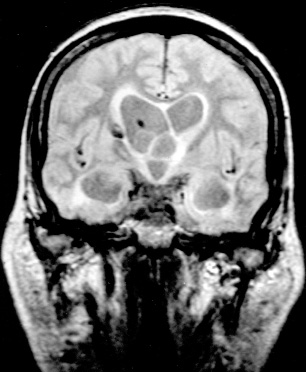
sional inflammatory reaction (Fig. 4), if they are ad-herent to the ventricular wall. In these cases, theependymal cell layer is altered, forming subependy-
Clinical Manifestations and Diagnosis
mal giant cells that tend to group and protrude into
With regard to its pathophysiology, neurocysticer-cosis may present as a high- or low-intensity diseasethat may be located in the ventricles, parenchyma,
meninges, or mixed location. It may manifest as anacute, subacute, or chronic disease, in remission orexacerbation; it may have a simple or complicatedclinical evolution, be asymptomatic, symptomatic,or fatal. It may subside with or without treatment; itssymptoms may disappear for long periods of time orpersist until death. The onset of the symptoms maybe insidious or abrupt, and the clinical course isvariable and unpredictable. The prognosis is alwayssevere [2].
The clinical presentation may have an insidious
beginning and lead to death within a few minutes orin over 30 years' time. The polymorphous nature ofthe symptoms is characteristic of the disease and isdue to a series of factors such as the number, loca-tion, form, size, and stage of development of the
parasite, the nature of the parasite's action in thehost's organism, and the host's individual immuno-logical response [63-66]. In 1988, Zee et al. [84] re-ported that out of 46 patients with intraventricularcysts, 6 died of hydrocephalus a short time after ad-mission.
Diagnosis may be reached by appropriate clinical
observation that includesepidemiological aspects,clinical and neurological examination, along with de-tailed study of the CSF and imaging studies such ascomputed tomography (CT) and magnetic resonanceimaging (MRI).
Cutaneous cysticercosis may be associated with
neurocysticercosis in 65% of the cases. It usuallysuggests a benign neurocysticercosis. The most
Fig. 4. a Coronal and b sagittal FLAIR MRI showing intra-
common clinical symptoms of the disease are:
ventricular cysts and ependymal inflammatory reaction
epilepsy, intracranial hypertension, psychiatric
Chapter 17 • Hydrocephalus in Neurocysticercosis and Other Parasitic and Infectious Diseases
changes, meningitis, and meningoencephalitis. Oc-
that occurs when cysticerci die and disintegrate, caus-
casionally, headache is the only complaint. Al-
ing antigen release [54,56].
though the association of these symptoms is com-
Intraventricular cysticercosis occurs in 11%-17%
mon, isolated epilepsy seems to be the predominant
of the patients, being a potentially lethal form of
form of presentation. Partial seizures are the most
the disease [85]. The oncospheres probably reach
frequent; intractable seizures are rare. Intracranial
the ventricles through the choroid plexus. They
hypertension is usually severe and tends to occur in
may develop and float freely in the CSF or adhere
association with other symptoms, especially epilep-
to the ependyma by a granulomatous reaction. Oc-
sy. Its treatment is difficult, although once the acute
clusion of the aqueduct or the foramina of Luschka
phase has passed, these children frequently present
and Magendie may result in acute obstructive hy-
a good recovery. Sequelae may ensue in some cases.
drocephalus, sometimes associated with sudden
Behavioral disturbances such as aggressiveness, ag-
death. Nausea, vomiting, dizziness, headache,
itation, and irritability may also be seen. One
diplopia, syncope, and altered state of conscious-
should bear in mind the possibility of these chil-
ness are common manifestations of intraventricu-
dren developing a clinical picture of neurological
and psychological regression that, when associated
CSF analysis is one of the most important exami-
with epilepsy, resembles a degenerative disease of
nations in the diagnosis of neurocysticercosis, even
though the results may be normal in approximately
The analysis of the disease is the most important as-
20%-25% of the cases despite the presence of viable
pect of its treatment. Neurocysticercosis can be divid-
cysticerci. In these cases, an indirect approach to di-
ed into two major forms: active and inactive. In the ac-
agnosis is a trial drug therapy. In roughly 75% of the
tive forms of the disease one may have clinical mani-
cases, the immunodiagnosis is positive or, at least,
festations such as (1) arachnoiditis, (2) hydrocephalus
there are changes in one or more of the CSF parame-
due to meningeal inflammation, (3) parenchymal
ters. On the other hand, if the patient's condition
cysts, (4) cerebral infarction due to vasculitis, (5) mass
permits, close clinical observation with CSF exami-
effect due to large cysts, (6) intraventricular cysts, and
nation, CT, and/or MRI may lead to an unequivocal
(7) spinal cysts. The inactive forms present as (1)
diagnosis. The definitive diagnosis of neurocysticer-
parenchymal calcifications and (2) hydrocephalus due
cosis is based on immunodiagnosis in the CSF
to meningeal fibrosis.
and/or on lesions suggestive of this parasitic disease
Cerebral cysticercosis may be entirely asympto-
on CT or MRI. The changes in CSF that characterize
matic, being demonstrated only at autopsy. In the
the syndrome are lymphocytic pleocytosis, in-
symptomatic cases the neurological examination is
creased eosinophilic content, elevated total protein
normal in 25% of the total [22]. The initial symptoms
levels, hypoglycorrhachia, and positive immunolog-
that prompt the patient to seek medical help are
ical reactions in the CSF. Two or more positive tests
seizures, meningeal signs, visual disturbances,
increase the certainty of the diagnosis. The comple-
headaches, and vomiting. Epilepsy may be an isolat-
ment fixation test, one of the first tests utilized for
ed symptom for a long time, being called idiopathic
the diagnosis of this disease, is positive in 83% of the
epilepsy still to the present day. Neurocysticercosis
cases of active meningeal cysticercosis if there are
may lead to an intracranial hypertension syndrome,
inflammatory changes of CSF. Conversely, the test
with headache, vomiting, dizziness, and papilledema.
sensitivity is only 22% if CSF examination is normal.
Altered mental status may be a manifestation of the
Another immunoassay used nowadays is the en-
disease, and pure psychotic forms are found in 15% of
zyme-linked radioimmunoassay, which has a speci-
the cases. Cysticerci on the motor or sensory cortical
ficity of 95% and a sensitivity of 87% in cases of ac-
areas can cause seizures, usually generalized. Chron-
tive meningeal neurocysticercosis.
ic focal epilepsy is related to residual calcifications
Images made by CT scanning vary according to
and areas of gliosis that represent the inactive form
the phase of maturation of the parasite and its loca-
of the disease.
tion. Viability of the cysticercus usually is deter-
Meningitis due to neurocysticercosis may show
mined by contrast enhancement. Neurocysticerco-
acute or chronic headaches, neck pain, and occasion-
sis may show on CT as single, multiple, or racemose
ally fever. In some cases, the typical presentation of
vesicles, generally localized in the brain parenchy-
increased intracranial pressure can be seen. In other
ma. The cysts have well-defined contours, no perile-
cases, meningitis may progress slowly and with only
sional edema, and little or no enhancement after
mild symptoms, leading to communicating hydro-
contrast administration; colloidal-phase cysts show
cephalus due to basilar arachnoiditis. The pathogene-
perilesional edema, have less defined contours, and
sis of neurocysticercosis is that of a chronic inflam-
may present annular contrast enhancement sepa-
matory process with an irregular period of activation
rating the cyst from surrounding cerebral edema.
This tomographic picture was defined as the acute
encephalitic phase. Granulomas indicate a degener-ating parasite; they may be single or multiple, ordi-narily nodular and located on the parenchyma. Cal-
It is known that neurocysticercosis may be treated
cifications are the most common tomographic find-
surgically or medically. Surgery for removal of cys-
ings. They present as minuscule hyperdense lesions
ticerci, employed in only a small and specific num-
not surrounded by edema, that do not change after
ber of cases (racemose cisternal cysticerci, ventricu-
administration of intravenous contrast; they may
lar cysticerci), is practically never used in children
be single or multiple, of various sizes, always round,
as these forms of the disease do not ordinarily occur
and appear at least 36 months after the start of the
in this age group. Carbamazepine is suggested for
degeneration. The presence of diffuse or localized
the control of epilepsy because of the increased fre-
edema without vesicles in the treated patient may
quency of partial seizures. Del Brutto and Sotelo
indicate the beginning of the evolutive phase, due to
[20-22] demonstrated that 83% of the patients were
an inflammatory reaction, hydrocephalus, in-
drug-free if anticonvulsants were used in conjunc-
creased ventricular size, sometimes in the absence
tion with cysticercocidal drug therapy. Conversely,
of vesicles, calcifications or granulomas. This is the
only 26% of the patients were drug-free when only
characteristic pattern of cysticercotic encephalitis,
anticonvulsants were utilized. Increased intracra-
a particularly severe form of neurocysticercosis in
nial pressure is treated specially with dexametha-
which the immune system responds actively and in-
sone, for a long period, 1 month or more, to with-
tensely against a massive invasion of the brain
draw slowly. For those few patients who present se-
parenchyma by cysticerci.
vere intracranial hypertension that is unresponsive
CT findings of subarachnoid neurocysticercosis
to dexamethasone, mannitol or a lumboperitoneal
include hydrocephalus, abnormal enhancement of
shunts might be used as a last resort.
the basal meninges, and cerebral infarction. Ventric-
Etiological treatment involves drugs that pene-
ular cysticerci may be isodense in relation to CSF,
trate the central nervous system and destroy the
making their visualization difficult on CT scan. Ven-
parasite. Currently two drugs are utilized: prazi-
triculography allows precise identification of these
quantel (PZQ) and albendazole (ABZ). Their use is
restricted to patients who exhibit intact forms of the
The MRI characteristics depend on the phase of
parasite, i.e., vesicles. The simultaneous administra-
the disease. Vesicular cysts are seen as round le-
tion of corticosteroids and cysticercocidal drugs to
sions with well-defined contours and a signal in-
patients with intraparenchymal lesions is also con-
tensity similar to CSF in both T1- and T2-weighed
troversial [20, 21]. ABZ has been the preferred initial
images. The scolex is seen as a hyperdense point
cysticercocidal drug, as it is cheaper, has fewer side
inside the cystic lesion. The details of the cyst wall
effects, and is used for a shorter period of time. If the
and perilesional edema, in addition to the pres-
lesions persist, PZQ may be used after 3 months, or
ence of intraventricular cysts, are better visual-
the ABZ treatment may be repeated [7, 16]. PZQ is an
ized. All MRI sequences can identify the vesicles.
isoquinoline with strong antiparasitic activity that
The degenerating vesicles are recognized by the
leads to the disappearance of 60%-70% of intra-
presence of edema, which appears bright on T2 se-
parenchymal cysts after 15 days of treatment at a
quences and is enhanced by contrast material.
dosage of 50 mg/kg per day. ABZ is a benzimidazole
Granulomas and residual calcifications appear as
with antihelmintic properties, currently the drug of
a signal void. These forms of neurocysticercosis
choice for the treatment of neurocysticercosis. It de-
demonstrate one of the most important diagnostic
stroys 75%-90% of intraparenchymal cysts when ad-
MRI. MRI easily diagnoses
ministered for 8 days at a dosage of 15 mg/kg per day.
meningeal cysticerci, as they present a signal dif-
Furthermore, it also acts on the meningeal and in-
ferent from the CSF.
traventricular forms of the disease, due to its good
In childhood, the diagnosis through imaging
penetration into the subarachnoid space [21]. De-
demonstrates two factors that are different from the
spite treatment with PZQ and ABZ, however, a large
disease in adults. The first is the number of lesions:
number of patients will require surgical treatment
only a small group presents with an intact parasite,
to treat hydrocephalus or remove isolated cysts
the vesicle; also, most images are from the acute phase
(Fig. 5). Patients presenting with compression of cra-
of the disease. Secondly, increased intracranial pres-
nial nerves, brain tissue, or spinal cord are also
sure in infancy is due to cerebral edema from the in-
amenable to surgery.
flammatory reaction and not to hydrocephalus sec-
Three mechanisms may produce increased in-
ondary to ventricular obstruction, the latter being
tracranial pressure: diffuse brain edema, hydro-
common in adult patients.
cephalus, or mass effect (pseudotumoral form).
Chapter 17 • Hydrocephalus in Neurocysticercosis and Other Parasitic and Infectious Diseases
complished. However, if the cysticerci lie in thecisterns and their degeneration process has al-ready started, complete removal may be difficultor even impossible.
Hydrocephalus can be identified in 15%-30% of thecases with neurological symptoms [42, 51]. in themajority of cases it derives from chronic basalarachnoiditis or meningeal fibrosis. A small per-centage of cases may be due to intraventricularcysts. It is believed that the larvae invade the ven-tricular system through the choroid plexus. Hydro-cephalus corresponds to 90.5% of the cases of in-tracranial hypertension treated with surgery. In-
volvement of the ventricular system is ordinarilyassociated with higher mortality and morbidityrates than is the intraparenchymal form of the dis-ease [4, 7, 42, 60]. There is still no consensus in theliterature as to the best kind of treatment. On thewhole, there are three types of treatment: drug ther-apy, which has little or no efficacy in this form of thedisease; ventricular shunts; and surgical removal ofcysts (Fig. 6).
Hydrocephalus may be produced by mechanical
obstruction of the CSF pathways by cysts (Fig. 7), in-flammatory reaction caused by cyst degeneration, oran association of both factors. Migration of the cystswithin the ventricles may lead to the intermittentheadaches that characterize Bruns syndrome. Many
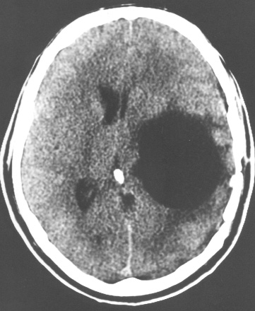
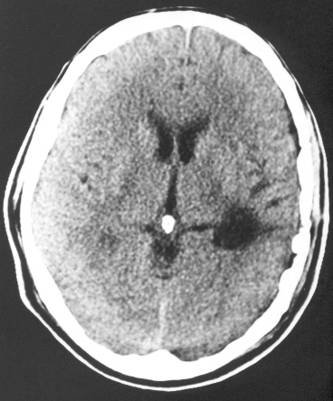
Fig. 5 a, b. Axial CT scans. a Blockage of temporal horn by
authors recommend ventricular shunts as the first or
ependymitis. a Postoperative follow-up after endoscopy
definitive form of treatment [7, 14, 15].
Ventriculoperitoneal shunts are the treatment of
choice for those patients who present with inflamma-tory obstruction of the ventricular system and hydro-cephalus. As a rule, the patients exhibit a good recov-
Diffuse Brain Edema
ery. However, mechanical and inflammatory compli-
This form is found in 2.8% of the cases that presentincreased intracranial pressure. It is seen in cases ofextensive infestation, cysts, inflammatory reaction,
and diffuse brain expansion.Treatment is mostly clin-ical, but in intractable cases lumboperitoneal shuntsare indicated. In extreme cases, decompressive cran-iotomies may be performed in a heroic attempt tosave lives.
Giant cysts may be located in the brain parenchy-ma or in the cisterns and are easily identified byimaging studies. If located within the parenchy-ma, complete removal of the cysts is readily ac-
Fig. 6. Treatment algorithm for patients with hydrocephalus
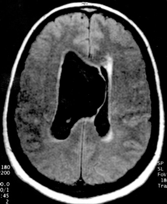
Fig. 7. a FLAIR MRI revealing a giant intraventricular cys-
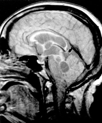
Fig. 8. Axial enhanced T1-weighted MRI. Clusters of cys-
ticercus. b Postoperative T1-weighted MRI. c Cysticercus
ticerci arising from the sylvian fissure. Note the normal ven-
cellulosae endoscopically extracted
tricular size. b Axial unenhanced T1-weighted MRI,postop-
erative follow-up after endoscopy
cations are prevalent among these patients, with a
Pitfalls in Surgical Treatment
high incidence of ventriculitis, meningitis, and ob-struction of the catheter by the cyst. Colli et al. [16] re-
Viable intraventricular cysts may shift position fre-
port an 82% (46 cases) shunt revision rate in a study
quently, moving from the lateral ventricles to the third
of 56 patients with neurocysticercosis treated by ven-tricular shunts. In those cases in which ependymitis
and fourth ventricles. A recent imaging study, prefer-
caused ventricular loculation, an endoscopic proce-
ably one obtained during the immediate preoperative
dure followed by a ventricular shunt showed good re-
period, should allow detection of any cyst migration
sults. Free intraventricular cysts, even those located
and thus help with the surgical strategy, hopefully pre-
inside the third and fourth ventricles and basal cis-
venting cysts from being overlooked during surgery.
terns, are easily removed by endoscopic procedures.
Neurocysticercosis is a chronic inflammatory diseasein which there may be communicating and noncom-
municating hydrocephalus (Fig. 6).
Cisternal forms with cranial nerve compression
It is estimated that 2.4 million people throughout the
(Fig. 8) and spinal forms with spine compression have
world have cysts from neurocysticercosis in the ven-
been described.
tricular cavities [42,70]. The cysts' lack of vasculariza-
Chapter 17 • Hydrocephalus in Neurocysticercosis and Other Parasitic and Infectious Diseases
tion and mobility allow easy handling and easy re-
intraventricular cysts [4]. The advantages of neuroen-
moval in the absence of ependymitis. Large cysts with
doscopy are numerous in comparison to craniotomy.
scolex in their interior need to be completely re-
Neal in 1995 [40] reported the removal of a cyst of the
moved. Even if the cyst ruptures during the proce-
posterior portion of the third ventricle, using a com-
dure, there is no associated ventriculitis. In the event
bination of rigid and flexible endoscope. Irrigation
of ependymitis with multiple intraventricular locula-
using positive pressure may be another tool to help
tions, endoscopy may allow communication between
mobilize and aspirate the cysts. In our experience, out
various cavities and placement of a single ventricular
of 310 patients who underwent endoscopic proce-
shunt system. Endoscopic third ventriculostomy is
dures, in 20 cases (7.4%) it was for treatment of hydro-
typically performed when there is a noninflammato-
cephalus associated with the disease. Bergsneider et
ry CSF blockage at the level of the aqueduct or fourth
al. [9], while studying ten patients with hydro-
ventricle (Fig. 6). Once an inflammatory process at
cephalus due to neurocysticercosis, concluded that
the level of the basal cisterns has occurred, its useful-
endoscopic procedures should be considered as the
ness is limited. In cases presenting the parenchymal
first treatment option. Only three of their patients re-
tumoral form of the disease, endoscopy is useful in
quired a shunt afterwards.
the removal of cysts and verification of other cystsnot detected by imaging studies. In the severe race-mose form, in which numerous cysts located within
Outcome of Hydrocephalus Due to
the interpeduncular cistern may present mass effect
and symptoms such as altered consciousness, visualand vegetative changes, endoscopic opening of the tu-
Given that hydrocephalus in neurocysticercosis can
ber cinereum can allow removal of these cysts and
be attributed to various mechanisms, it may be said
improvement of the symptoms.
that the outcome will depend on its etiology. Intra-
Surgical approach to the lateral or third ventricles
ventricular cysts can be treated using endoscopic
through a transcallosal or transcortical-transventric-
techniques or a direct approach, as for cysts of the
ular approaches is recommended for the treatment of
fourth ventricle (Fig. 9) or of the periaqueductal re-
Fig.9. a,b Axial and sagittal T1-weighted MRI.Fourth ventricular cysticercus and scolex.c,d Endoscopic views of the floor of
the third ventricle. MB, mammillary bodies; Aq, cerebral aqueduct; PC, posterior commissure. Note the cyst appearing inside
the fourth ventricle. e Endoscopic view of the cysticercus being extracted. f Sagittal T1-weighted MRI, 3-month follow-up
ing human tapeworm infection would be an alter-native control technique. The economic cost oftreating a case of tapeworm intestinal infection is150 times smaller than the cost of the same medica-tion used to treat neurocysticercosis [47]. At thepresent time there are two kinds of strategies rec-ommended by the World Health Organization: thefirst, for short term results, is based on widespreadtreatment of tapeworm infection and foci of trans-mission; the other, aimed at long-term results, fo-
cuses on the development of breeding and inspec-tion techniques for pork, adequate sanitary mea-
Fig. 10. Sagittal T1-weighted MRI. Mesencephalic hydatic
sures, and actions to detect and treat humans
infected by tapeworms [47].
gion (Fig. 10). Communicating hydrocephalus result-ing from inflammatory blockage of the basal cisterns
Other Parasitic Diseases Responsible
and treated by ventricular shunts (Fig. 6) may presenta higher incidence of complications [13]. Recurrent
inflammatory reactions with CSF pleocytosis and in-creased protein levels due to the rupture of small in-traventricular cysts is probably one of the causes of
these complications [13]. It is known that drug thera-py for neurocysticercosis is far from ideal and for the
Histoplasma capsulatum is a dimorphic fungus that
intraventricular forms of the disease is not efficacious
exists as a mycelial form at ambient temperature and
at all [63]. In a series that studied large groups of pa-
as a yeast form in mammals. Its distribution is univer-
tients, forms of neurocysticercosis considered hyper-
sal, being endemic in certain regions of the United
tensive comprised 20%-36% of the cases [48, 59, 66]
States and Latin America [75]. The fungus exists in the
and usually resulted in the worst outcome. Stepien
soil of endemic areas, especially in dusty places where
[59] reported on 43 patients who presented hydro-
the soil contains bat or bird feces, such as for instance
cephalus. There was improvement in only 12 cases
a hen house [30]. The initial infection is due to spore
(28%); 29 patients died (67%). In a study from the city
inhalation. In endemic areas, almost the entire popu-
of Ribeirão Preto (Brazil) of 500 patients in a period
lation is infected and subject to multiple cases of re-
of 23 years [66], 68 (13.6%) underwent some form of
infection [72, 76].
CSF shunt diversion or surgical removal of cysts. Out
Disseminated chronic histoplasmosis is a rare
of 74 patients classified as having a pure hypertensive
event, its incidence estimated at 1 per 100 000 to 1 per
form of the disease, 21 (28.4%) died. Canelas [14] not-
150 000 infected individuals per year [30]. Sympto-
ed that of 63 patients who underwent a surgical pro-
matic CNS involvement is believed to occur in 10%-
cedure, 28(44.4%) died. Takayanagui [64] pointed out
25% of the cases of disseminated histoplasmosis [18,
that, of his series of 56 patients that presented with
45, 49, 74].
signs of increased intracranial pressure, 12 (21.4%)
Shapiro classified the lesions of histoplasmosis of
died and 12 developed incapacitating neurological se-
the CNS as follows: (1) miliary granuloma; (2) histo-
quelae. Studies concerning the intellectual develop-
plasmoma; (3) meningitis/ventriculitis, the most fre-
ment of these patients are extremely difficult, as
quent CNS presentation, affecting preferentially the
pointed out by Scharf [48], for each patient has a dif-
skull base meninges; (4) spinal compression [30, 50].
ferent individual outcome depending on the lesions,
The diagnosis of histoplasmosis is becoming more
the number of parasites, the duration of the infesta-
and more common due to an increase in the number
tion, and the immunological response of the host.
of immunocompromised patients due to AIDS or im-munosuppression.Ventriculitis can progress to hydro-cephalus, requiring a CSF shunt. In most cases, many
shunt revisions are necessary. In other cases, a
parenchymal lesion is detected and a biopsy suggest-
Control of neurocysticercosis demands attention
ed. Histoplasma capsulatum should be considered in
to hygiene habits and environmental sanitation.
the differential diagnosis even in cases of communi-
Widespread medical treatment aimed at eradicat-
cating idiopathic hydrocephalus (no associated
Chapter 17 • Hydrocephalus in Neurocysticercosis and Other Parasitic and Infectious Diseases
meningitis or ventriculitis). A common factor among
3. Agapajev S, Alves-Moreira D, Barriviera B. Neurocysticer-
all cases, irrespective of the clinical presentation, is the
cosis:treatment with albendazole and destrocloropheri-
insidious clinical course, characterized by numerous
nami. Trans R Soc Trop Med Hyg 83:377-83, 1989
admissions to the hospital, a number of times because
4. Apuzzo MLJ, Dobukin WR, Zee C, et al. Surgical consider-
ations in treatment of intraventricular cysticercosis. An
of a shunt revision, with no etiological diagnosis.
analysis of 45 cases. J Neurosurg 60:400-407, 1984
The authors have treated eight cases of histoplas-
5. Aristotle. Historie des Animaux, vol III, book VIII, para XXI.
mosis, one being in a 12-year-old patient who had al-
Paris, Societé d'Editions Les Belles Lettres, pp 48-49, 1969
ready undergone 28 shunt revisions and an erroneous
6. Bamberger DM. Successful treatment of multiple cerebral
tuberculosis treatment. Another patient had had 16
histoplasmomas with itraconazol. CID 28:915-916, 1999
shunt revisions. After treatment the shunt complica-
7. Bandres JC, White AC, Samo T, Murphy EC, Harris RL. Ex-
tions disappeared. One patient underwent an endo-
traparenchymal neurocysticercosis: report of five casesand review of management. Clin Infect Dis 15:799-81, 1992
scopic third ventriculostomy that obliterated 3
8. Bérard H, Astoul PH, Frenay C, et al. Histoplasma dis-
months later.
séminée à Histoplasma capsulatum avec atteinte cérébrale
Another common characteristic shared by these
survenue 13 ans après la primo-infection. Ver Mal Resp
cases are the CSF findings. They are characterized by
16:829-831, 1999.
a modest increase in cell number, usually below ten
9. Bergsneider M,Holly LT,Lee JH,King WA,Frazee JG.Endo-
cells, mostly due to lymphocytic/monocytic infiltrate,
scopic management of cysticercal cysts within the lateral
and modest hypoglycorrhachia or normal glucose
and third ventricles. Neurosurg Focus 6(4):article 7, 1999
levels; protein content is always related to the phase of
10. Boppana S, Pass RF, Britt WS. Symptomatic congenital cy-
tomegalovirus infection: neonatal morbidity and mortali-
the disease. During recurrence episodes, levels are
ty. Pediatr Infect Dis J 11:93-99, 1992
more elevated and protein/cell dissociation is more
11. Boudawara MZ, Jemel H, Ghorbel M, et al. Les kystes hyda-
evident. Intraventricular protein levels are usually
tiques du tronc cerebral. A propos de deux cas. Neu-
above 1 g and may continue to rise even if external
rochirurgie 4:321-324, 1999.
ventricular drainage is instituted.
12. Bruck I, Antoniuk SA, Wittig E, Accorsi A. Neurocisticer-
The rate of positive results obtained in CSF cultures
cose na infância: diagnóstico clínico e laboratorial. Arq
varies from 25%-to 65%. The best results can be ob-
Neuropsiquiatr 49:43-46, 1991
tained with bone marrow cultures, which have a posi-
13. Canelas HM. Neurocisticercose: incidência, diagnóstico e
formas clínicas. Arq Neuropsiquiatr 20:1-16, 1962
tivity index of about 75%. Blood cultures are positive
14. Canelas, HM. Cisticercose do sistema nervoso central. Rev
in 50%-70% of the cases [46]. The mean time for the
Hosp Clin Fac Med S Paulo 47:75-89, 1963
results of the cultures is 4 weeks. Serological test re-
15. Colli BC, Assirati JA, Machado HR, Santos F, Takayanagui
sults for histoplasmosis are very difficult to interpret.
OM. Cysticercosis of the central nervous system II. Spinal
Antibody detection tests in the CSF have a positivity of
cysticercosis. Arq Neuropsiquiatr 52:187-199, 1994
about 80% and antibody detection in the blood a pos-
16. Colli BO,Martelli N,Assirati JA,et al.Cysticercosis of the cen-
itivity rate of 92%. However, the rate of false-positive
tral nervous system I.Arq Neuropsiquiatr 52:166-186, 1994
17. Colli BO, Martelli N, Assirati JA, et al. Results of surgical
results must also be considered. Cross-reaction rates
treatment of neurocysticercosis in 69 cases. J Neurosurg
can reach 50%, especially with tuberculosis and other
fungal infections (mainly aspergillosis, blastomycosis,
18. Cooper RA, Golstein E. Histoplasmosis of the central ner-
coccidioidomycosis) [73]. In 1986, Wheat et al. [77]
vous system. Report of two cases and review of the litera-
proposed studying the histoplasma polysaccharide
ture. Am J Med 35:45-47, 1963
antigen (HPA) in blood, urine, and CSF. The best posi-
19. Davis LE. Fungal infections of the central nervous system.
tivity rates (91%) are obtained in the urine of patients
Neurol Clin 4:761-781, 1999
with disseminated disease. When the disease is re-
20. Del Brutto OH. Cisticercosis and cerebrovascular disease:
a review. J Neurol Neurosurg Psychiatry 55:252-254, 1992
stricted to the CNS,positivity falls to 19%.The classical
21. Del Brutto OH, Sotelo J. Albendazole therapy for sub-
treatment consists of intravenous amphotericin B for 3
arachnoid and ventricular cisticercosis: case report. J
weeks and fluconazole for 6 months during the main-
Neurosurg 72:816-817, 1990
tenance phase of the treatment. Once treatment is ini-
22. Del Brutto OH, Sotelo J. Neurocysticercosis: an update.
tiated, there is a rapid fall in CSF protein levels, reduc-
Rev Infect Dis 10:1075-1087, 1988
ing shunt obstruction incidents.
23. Enarson DA, Keys TF, Onofrio BM. Central nervous sys-
tem histoplasmosis with hydrocephalus. Am J Med64:895-896, 1978
24. Escobar A. The pathology of neurocysticercosis. In: Pala-
cios E, Rodrigues-Carbajal J, Taveras (eds) Cysticercosis of
1. Abbassioun K, Amirjamshidi A, Moinipoor MT. Hydatic
the central nervous system. Charles C. Thomas, Spring-
cyst of the pons. Surg Neurol 26:297-300, 1986
field, pp 27-54, 1983
2. Acha PN, Szyfres B. Zoonosis enfermedades transmisibles
25. Estanol B, Kleriga E, Loyo M, et al. Mechanisms of hydro-
communes de hombre y a los animals. OMS 503:763-774,
cephalus in cerebral cysticercosis: implications for thera-
py. Neurosurgery 13:119-123, 1983
26. Flisser A, Woodhouse H, Larralde L. Human cysticercosis;
50. Shapiro JL, Lux JJ, Sprofkin BE. Histoplasmosis of the cen-
antigens, antibodies and nonresponders. Clin Exp Im-
tral nervous system. Am J Pathol 31:319-334, 1955
munol 39:27-31, 1980
51. Sotelo J, Marin C. Hydrocephalus secondary to cysticer-
27. Frenkel, JK. Toxoplasmosis: mechanisms of infection, lab-
cotic arachnoiditis. A long term follow-up review of 92
oratory diagnosis and management. Curr Top Pathol
cases. J Neurosurg 66:686-689, 1987
52. Sotelo J, Escobedo F, Rodrigues J, Torres B, Rubio F. Thera-
28. Frenkel JK, Friedlander S. Toxoplasmosis. Pathology of
py of parenchymal brain cysticercosis with praziquantel.
neonatal disease. Pathogenesis, diagnosis and treatment.
N Engl J Med 310:1001-1007, 1984
Public Health Service Publication no 141. US Government
53. Sotelo J, Guerrero V, Rubio F. Neurocysticercosis: a new
Printing Office, Washington, DC, 108 pp, 1952
classification based on active and inactive forms. A study
29. Go JL, Kim PE, Ahmadi J, Segall HD, Zee CS. Fungal infec-
of 753 cases. Arch Intern Med 145:442-445, 1985
tions of the central nervous system. Neuroimaging Clin
54. Spina- França A. Aspectos biológicos da neurocisticer-
North Am 10:409-425, 2000
cose: alterações do liquido cefalorraquidiano. Arq Neu-
30. Goodwin RA, Shapiro JL, Thurman GH, Thurman SS, Des
ropsiquiatr São Paulo 20:17-30, 1962
Prez M. Disseminated histoplasmosis: clinical and patho-
55. Spina-França A, Livramento J A , Machado LR. Cysticerco-
logical correlations. Medicine 59:1-33, 1980
sis of the central nervous system and cerebral fluid: im-
31. Gottlieb T, Marriott D. Disseminated histoplasmosis in an
munodiagnosis of 1573 patients in 63 years (1929-1992).
AIDS patient. Aust N Z Med 20:621-622, 1990
Arq Neuropsiquiatr 51:16-20, 1993
32. Hanshaw JB, Dudgeon JA (eds) Viral diseases of the fetus
56. Spina-França A. Neurocisticercose e imunologia. J Bras
and newborn. Saunders, Philadelphia, 1978
33. Karalakulasingam R, Arora KK, Adams G, Serratoni F,
57. Spina-França A Incidência de neurocisticercose no
Martin DG. Meningoencephalitis caused by Histoplasma
serviço de neurologia do Hosp. Das Clinicas da FMUSP.
capsulatum. Arch Intern Med 136:217-220, 1976.
Rev Paul Med 43:160-161, 1953
34. Khamlich A, Belefkih N, Guarzazi A, Chkili T, Bellakhdar
58. Stagno S, Reynolds DW, Huang ES. Congenital cy-
F. Le kyste hydatique de la fossa cerebrale postérieure (Re-
tomegalovirus infection: occurrence in an immune popu-
vue de la literature à propos de 5 cas). Maroc Med 4:223-
lation. N Engl J Med 296:1254-1258, 1977
59. Stepién L Cerebral cysticercosis in Poland: clinical symp-
35. Knapp S, Turnherr M, Dekan G, et al.A case of HIV-associ-
toms and operative results in 132 cases. J Neurosurg
ated cerebral histoplasmosis succesfully treated with flu-
conazole. Eur J Clin Microbiol Infect Dis 88:658-661, 1999
60. Stern WE. Neurosurgical considerations of cysticercosis
36. Lambert RS, George RB. Evaluation of enzyme im-
of the central nervous system. J Neurosurg 55:382-385, 1981
munoassay as a rapid screening test for histoplasmosis
61. Sullivan AA, Benson SM, Ewart AH, et al. Cerebral histo-
and blastomycosis. Am Rev Respir Dis 136:316-319, 1987
plasmosis in an Australian patient with systemic lupus
37. LeBourgeois PA. Isolated central nervous system histo-
erythematosus. MJA 169:201-202, 1998
plasmosis. Southern Med J 72:1624-1625, 1979
62. Tabbal SD, Harik SI. Cerebral histoplasmosis. N Engl J
38. Lombardo L, Mateos JH, Estanol B. La cisticercosis cere-
Med 15:1176, 1999
bral en México. Gac Med Mex 118:1-8, 1982
63. Takayanagui OM. Neurocisticercose II. Avaliação da ter-
39. Mitchel WG, Crawford TO. Intraparenchymal cerebral
apeutica com praziquantel.Arq Neuropsiquiatr São Paulo,
cysticercosis in children: diagnosis and treatment. Pedi-
48:11-15, 1990.
atrics 82:76-82, 1988
64. Takayanagui OM, Castro e Silva AA, Santiago RC,
40. Neal JH.An endoscopic approach to cysticercosis cysts of the
Odashima NS, Terra VC, Takayanagui AMM Notificação
posterior third ventricle. Neurosurgery 36:1040-1043, 1995
compulsória da cisticercose em Ribeirão Preto- SP. Arch
41. O'Doherty DS: Invasion of central nervous system by cys-
Neuropsiquiatr 54:557-564, 1996
ticercosis cellulosae. Georgetown Med Bull 15:128-136, 1961
65. Takayanagui OM, Jardim E.Therapy for neurocysticerco-
42. Obrador S. Cysticercosis cerebri. Acta Neurochir 10:320-
sis: comparison between albendazole and praziquantel.
Arch Neurol 49:290-294, 1994
43. Pratt-Thomas HR, Cannon WM. Systemic infantile toxo-
66. Takayanagui OM, Jardim E Aspectos clínicos da neurocis-
plasmosis. Am J Pathol 22:779-795, 1946
ticercose – análise de 500 casos. Arq Neuropsiquiatr São
44. Rawlinson WD, Packham DR, Gardner FJ, MacLeod C.
Paulo 41:50-63, 1983
Histoplasmosis in the acquired immunodeficiency syn-
67. Tiraboshi I, Parera IC, Pikielny R, Scattini G, Micheli F.
drome (AIDS). Aust N Z J Med 19:707-709, 1989
Chronic Histoplasma capsulatum infection of the central
45. Salaki JS, Louria DB, Chmel H. Fungal and yeast infections
nervous system siccessfully treated with fluconazole. Eur
of the central nervous system. Medicine 63:108-132, 1984
Neurol 32:70-73, 1992
46. Sathapatayavongs B, Batteiger BE,Wheat J, Slama TG,Wass
68. Tynes BS, Crutcher JC, Utz JP. Histoplasma meningitis.
JL. Clinical and laboratory features of disseminated histo-
Ann Intern Med 59:615-618, 1963
plasmosis during two outbreaks. Medicine 62:263-70, 1983
69. Walpole HT, Gregory DW. Cerebral histoplasmosis. South-
47. Schantz PM. Echinococosiasis (hydatidosis). In: Warren
ern Med J 80:1575-1577, 1987
KS, Mahmound AAF (eds) Tropical and geographical
70. Wei G, Li C, Meng J, Ding M. Cysticercosis of the central
medicine. McGraw Hill, New York, pp 487-497, 1984
nervous system. A clinical study of 1400 cases. Chinese
48. Scharf, D. Neurocysticercosis. Two hundred thirty-eight cas-
Med J 101:493-500, 1988
es from a California hospital.Arch Neurol 45: 777-780, 1988
71. Weller TH. The cytomegaloviruses: ubiquitous agents
49. Schulz DM. Histoplasmosis of the central nervous system.
with protean clinical manifestations. N Engl J Med
JAMA 151:549-551, 1953
285:203-214, 1971
Chapter 17 • Hydrocephalus in Neurocysticercosis and Other Parasitic and Infectious Diseases
72. Wheat J, French MLV, Batteiger B, Kohler R. Cerebrospinal
79. Wilson CB, Remington JS. What can be done to prevent
fluid histoplasma antibodies in central nervous system
congenital toxoplasmosis? Am J Obstet Gynecol 138:357-
histoplasmosis. Arch Intern Med 145:1237-1240, 1985
73. Wheat J, French MLV, Kamel S, Tewari RP. Evaluation of
80. Wolf A, Cowen D. Perinatal infections of the nervous cen-
cross-reaction in Histoplasma capsulatum serologic tests.
tral system. J Neuropathol Exp Neurol 18:191-243, 1959
J Clin Microbiol 23:493-499, 1986
81. Wolf A, Cowen D, Paige BH. Toxoplasmic en-
74. Wheat J. Histoplasmosis – experience during outbreaks in
cephalomyelitis. VI. Clinical diagnosis of infantile or con-
Indianapolis and review of the literature. Medicine
genital toxoplasmosis. Survival beyond infancy. Arch
Neurol Psychiatr 48:689-739, 1942
75. Wheat LJ, Batteiger BE, Sathapatayavongs B. Histoplasma
82. Wong SY, Remington JS. Toxoplasmosis in the setting of
capsulatum infections of the central nervous system – a
AIDS. In: Broder S, Merigan TC, Bolognesi D (eds). Text-
clinical review. Medicine 69:244-260, 1990
book of AIDS medicine. Williams & Wilkins, Baltimore,
76. Wheat LJ, Kohler RB, Tewari RP, Garten M, French MLV.
Significance of Histoplasma antigen in the cerebrospinal
83. Young RF, Gade G, Grinnell V. Surgical treatment for fun-
fluid of patients with meningitis. Arch Intern Med
gal infections in the central nervous system. J Neurosurg
149:302-304, 1989
77. Wheat LJ, Kohler RB, Tewari RP. Diagnosis of disseminat-
84. Zee C, Segall HD, Boswell W. MR imaging of neurocys-
ed histoplasmosis by detection of Histoplasma capsula-
ticercosis. J Comput Assist Tomogr 12:927-34, 1988
tum antigen in serum and urine specimens. N Engl J Med
85. Zee CS, Segal HD, Miller C, et al. Unusual neuroradiologi-
cal features of intracranial cysticercosis. Radiology
78. White HH, Fritzlen TJ. Cerebral granuloma caused by
137:397-407, 1980
Histoplasma capsulatum. J Neurosurg 19:260-264, 1962
Please confirm this as corresponding author.
Please confirm you mean the whole of southern Africa, not just South Africa. "Natives" is a bit of a
problematic word - it would be better to find an alternative. Are you referring to all poor black popu-
lations, whether urban or not, or just those living a more traditional rural life outside the towns?
"Usually people are infected by eating contaminated pork; however, immune-suppressed persons may
become infected by contaminated pork" - presumably there is supposed to be a difference, since you
say ‘however' instead of ‘and', but what is it? Please clarify.
Please confirm "tissue" for "tecidual", which does not exist in English. Occurs again.
Please confirm "phase".
If you are giving anticonvulsant and cysticercocidal drug therapy, the patients cannot be said to be
drug-free. What are you intending to say here? That they became drug-free, rather than having chron-
ic drug treatment?
"To withdraw slowly" - does this mean "in order for the ICP to go down slowly", or does it mean the
dexamethasone is slowly tapered off at the end of treatment?
"Despite treatment with" replaces "despite the use of ". The sentence now suggests that patients may
have to undergo surgery despite having been treated with the drugs, rather than suggesting that, al-
though some patients can be treated with the drugs, others have to undergo surgery. Please confirm
the change is correct.
What is "clinical"? Medical?
[Q 10] Please confirm this is what was meant by "positivity".
[Q 11] Please give an abbreviated form of this journal title in accordance with Index Medicus style.
[Q 12] Same again, please.
[Q 13] Same again, please.
[Q 14] Please confirm legend for Fig. 6 (treatment algorithm).
Source: http://www.clinicazymberg.com.br/pdf/capitulocisti.pdf
Microsoft word - cómo hacer más eficiente departamento auditoría interna corerif 2013
¿Cómo hacer más eficiente el Departamento de Auditoría Interna bajo un ENFOQUE DE RIESGOS? Nahun Frett, MBA, CIA, CCSA, CRMA, CFE, CPA Vicepresidente Auditoría Interna Central Romana Corporation, Ltd. República Dominicana Contenido Presentación: ¿Cómo hacer más eficiente el Departamento de Auditoría Interna bajo un Enfoque de Riesgos?
Microsoft word - challenge 8 page.doc
INTERNATIONAL LAW - RISK MANAGEMENT - EFFECTIVE POLICING CRIMINAL JUSTICE A Public Discussion Document Don Barnard & Alun Buffry, BSc. Dip Com (Open) The outcome of the cannabis debate cannot be predicted, but changes to the law must not take place without the most careful consideration of all the issues. You have a legitimate and important role to play - but only if you are willing to ask and address tough