Presentación de powerpoint


EuroResidue VIII – 23 -25 May 2016 - Egmond aan Zee, The Netherlands
Nora Mestorino, Martín Daniele, Martín Dadé, Andrea Buchamer, Valeria Vedovato, María Laura Marchetti
Laboratory of Pharmacological and Toxicological Studies (LEFyT), Faculty of Veterinary Science, Universidad Nacional de La Plata, 60 and 118, 1900 La Plata,
Doxycycline (DOX) is a semi-synthetic bacteriostatic tetracycline and a broad-spectrum antibiotic 1,2.
Although there are few pharmacokinetic studies in chickens, it is frequently used for the treatment of colibacillosis, salmonellosis, staphylococcal infections, avian mycoplasmosis and chlamydiasis1.
The misuse of DOX, illegal administration to animals, overdosing, and withdrawal period failure can lead to accumulation of residues of this antibiotic in edible animal tissues. Due to persistence of DOX residues in tissues and for consumer's health protection, the European Commission had set maximum residue limits (MRLs) for this compound at 100 μg/kg in muscle, 300 μg/kg in the liver and skin/fat, and 600 μg/kg in the kidney.
According to Commission Regulation (EU 37/2010) and EMA Summary Report Doxycycline 2, quantification of DOX in animal tissues requires a determination of only the parent compound as the residue marker, without its 4-epimer.
The antibiotics residue levels reached in organs and the rate of their depletion from tissues depend on the method of administration, animal species, as well as dose and specific given drug3. The differences in the antibiotic concentration and time of its depletion may be also influenced by the differences in the intake of drinking water by animals.
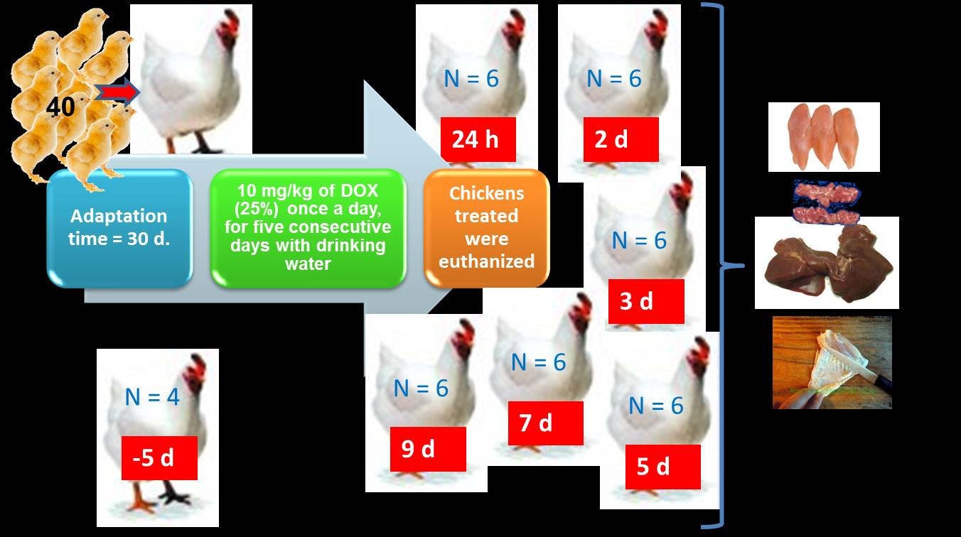
The purpose of the present study was to evaluate the withdrawal time (WT) of DOX formulation at 25 % in edible tissues, after its PO use in broilers.
Withdrawal time analysis
Study design treatment and samples colleted
2- 0.4 g of tissue + Mc
Ilvaine- EDTA (1.4 mL)
5-be centrifuged
1- Grinding the tissue
Supernatant S1
Doxycycline concentrations in function of time
found in muscle, kidney, liver and skin/fat were
6- S1+ S2 + S3
+ S4
plotted and analysed with the program WT
9-HPLC ASSAY
version 1.4 in order to recommend a period of
withdrawal time for this experimental
C column (Luna, 5 µm, 4.6 mm x 150 mm;
Phenomenex, Torrance,CA, USA) was eluted with
water-acetonitrile with 0.02 M oxalic acid and 0.0005M
7- SPE extraction
EDTA (72:28, v/v) at 1.2 mL/ min flow rate UV-VIS detector (346 nm)
This method performed accurately and reproducibly over a range of 0.1 to 6 µg/mL or µg/g for DOX. A linear relationship was obtained across one dynamic range (r values ranged from 0.9923 to 0.9997) (Fig. 1).
No matrix interferences were observed on the chromatograms of the samples with the same retention time as DOX. The chromatographic analysis time was short and DOX was presented in 4 min as a sharp and symmetrical peak with no interfering peaks (Fig 2).
The limits of detections (LODs) were 0.040, 0.024, 0.024 and 0.010µg/g and the LOQs were 0.095, 0.074, 0.054 and 0.100µg/g for kidney, skin/fat, muscle and liver respectively. The inter-day and intra-day variation in accuracy (recovery) and precision are presented in Table 1.
DOX concentrations were determined in all tissues examined; generally falling below the MRL at 7 d after administration is terminated. The mean ± SD DOX tissue concentrations after the PO administration of doxycycline to chickens in muscle, liver, kidney and skin/fat tissues are presented in Figure 3.
The maximum determined concentration in the kidney was 6.5μg/g and in the liver was 4.79 μg/g. Whereas in muscle and skin/fat the detected values were lower, with maximum concentration of 1.46 μg/g and 1.23 μg/g, respectively. At 7 d after treatment was completed, the DOX concentration in muscle was above LOQ.
The calculated withdrawal time was 6.58, 8.18, 8.69 and 6.96 d for muscle, liver, kidney and skin / fat, respectively (Figure 4 A, B, C and D).
Intra-day
Inter-day (over 3 days)
Fig4: Plot of the withdrawal times calculation for DOX in chicken
muscle (A), liver (B), kidney (C), skin/fat (D) and at the time
Fig 1. DOX calibration curve
when the one-sided 95% upper tolerance limit is below the EU
MRL for DOX after its oral administration (5 doses of 10 mg/kg
Fig3. Mean tissue concentrations of doxycycline in
body weight of DOX 25%). [Residue marker: doxycycline].
chickens slaughtered 1, 2, 3, 5, 7 and 9 d after oral
administration of DOX 25% (dose of 10 mg/kg body
weight during 5 days).
Area HPLC
Concentration (µg/mL)
Skin/Fat
Table 1. Validation results for the analysis of DOX in
chickens tissues
Fig 2. HPLC Chromatograms of DOX
The development and validation were successfully accomplished. This method performed accurately and reproducibly over a range of 0.1 to 6 µg/mL or µg/g for DOX.
The data obtained after DOX administration with water shows, that at the beginning after treatment, DOX reached high concentrations in all edible tissues. One day after administration of the last dose, DOX concentration rapidly decreased in all assayed tissues. Then, the residues decreased gradually and only trace concentrations were observed on day 9.
Our results demonstrate that DOX oral administration at the 10 mg/kg for five days together with drinking water requires withdrawal time respecting the MRL fixed for 9 days for human consumption.
1.- Anadón A., Martínez-Larrañaga MR., Díaz MJ., Bringas P., Fernández MC., Fernández-Cruz ML., Iturbe J., Martínez MA. Pharmacokinetics of doxycycline in broiler Chickens. Avian Pathology 1994; 23, 79-90 2.- Prats, C., Elkorchi G., Giralt M., Cristofol C., Pena J., Zorrilla I., Saborit J., Perez B. PK and PK/PD of doxycycline in drinking water after therapeutic use in pigs. J. Vet. Pharmacol. Ther., 2005; 28: 525-530. 3.- Kung K., Wanner M.: Pharmacokinetics of doxycycline in turkeys and comparison between feed and water medication. Archiv Geflugelkunde 1994, 58, 84–88.
Mr. Pedro Zeinsteger is acknowledged for check the manuscript.
Source: http://www.euroresidue.nl/images/posters2016/Poster%20112.pdf
eloslazer.com.tr
Journal of Cosmetic and Laser Therapy ISSN: 1476-4172 (Print) 1476-4180 (Online) Journal homepage: Successful removal of freckles with the bipolar radiofrequency and optical energy To cite this article: Osman Köse (2016) Successful removal of freckles with the bipolar radiofrequency and optical energy, Journal of Cosmetic and Laser Therapy, 18:4, 230-233, DOI: To link to this article:
Phytoprost:
Studien allgemein – Bedeutung in der Prävention von Krebs Cancer Causes Control. 2015 Nov;26(11):1521-50. doi: 10.1007/s10552-015-0659-4. Epub 2015 Sep 9. A systematic review of dietary, nutritional, and physical activity interventions for the prevention of prostate cancer progression and mortality. Hackshaw-McGeagh LE1,2, Perry RE3, Leach VA4,5, Qandil S4, Jeffreys M4, Martin RM3,4, Lane JA3,4.